Figure 5.
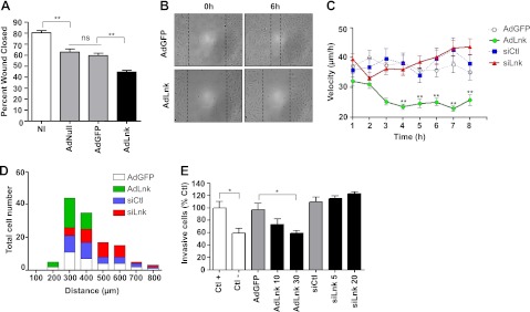
Lnk decreases EC motility in migration assays. A) ECs were NI or transduced with either control adenoviruses (AdNull, AdGFP) or AdLnk and subjected to a scratch assay. Wound closure was followed for 18 h by time-lapse imaging. Percentage of wound closed 6 h after wounding was measured and expressed as a percentage of wound closed compared to control (t=0 h, mean±se; n=4). B) Representative pictures of healing for AdGFP- vs. AdLnk-transduced EC monolayers at 0 and 6 h after wounding. C, D) ECs were transduced with AdGFP, AdLnk, control siRNA (siCtl), or Lnk-specific siRNA and seeded onto 10-μm-wide lines of CYTOO chips. Cell migration was monitored for 8 h. Cell velocity (C) and total covered distances (D) were quantified using MetaMorph imaging software (mean±se; n=60). E) ECs were not transduced (Ctl+ and Ctl−) or transduced with AdGFP (MOI 30), AdLnk (MOI 10 and 30), control siRNA (20 nM), or Lnk-specific siRNA (5 and 20 nM). After 48 h of silencing and 24 h of infection, HAECs were plated to the upper chamber, and 20% FCS was added in the bottom chamber as chemoattractant molecule, except in Ctl− conditions. After 18 h incubation, numbers of cells that migrated through the membrane were estimated using MTT colorimetric assay (mean±se; n=3). ns, not significant. *P < 0.05, **P < 0.01 vs. control.

