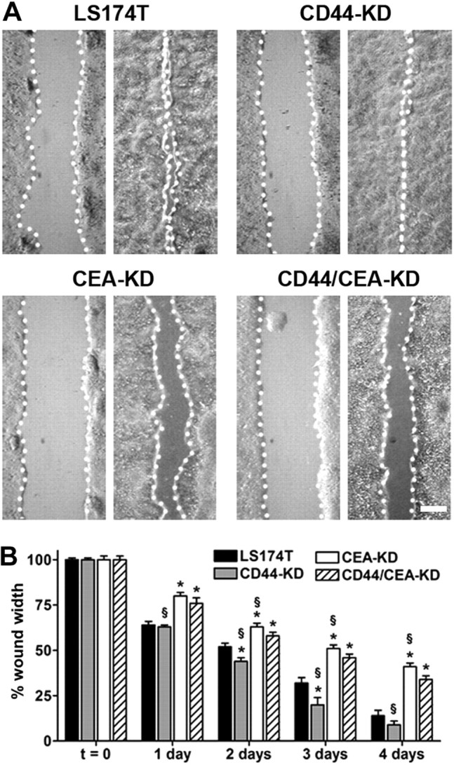
Figure 4.

CD44 knockdown augments, while CEA knockdown limits, wound healing. LS174T parental and CD44-KD, CEA-KD, and CD44/CEA-KD cells were detached and plated at 100% confluence; cells were allowed to attach to tissue culture-treated polystyrene for 6 h. Thin wounds were created and imaged (t=0), with subsequent images tracking wound closure taken at 24 h intervals up to 4 d. Initial wound widths for LS174T and CD44-KD, CEA-KD, and CD44/CEA-KD cells were 550 ± 60, 600 ± 6, 620 ± 10, and 570 ± 9 μm, respectively. A) Representative micrographs demonstrate initial (left panels) and final (right panels) wound widths. Scale bar = 200 μm. B) Bars indicate mean ± se wound widths for LS174T (solid bars), CD44-KD (shaded bars), CEA-KD (open bars), and CD44/CEA-KD (hatched bars). *P < 0.05 vs. LS174T cells; §P < 0.05 vs. CD44/CEA-KD cells.
