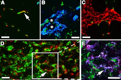
Figure 3.
PDGF-BB restores normal pericyte coverage to VEGF-induced vessels. Vessels induced by implantation of control CD8 (A), V (B), P (C), or VIPhigh myoblasts (D, E; n=3) in hindlimb tibialis anterior muscles were immunostained with antibodies against CD31 (endothelium, green), NG2 (pericytes, red), and αSMA (smooth muscle cells, blue in A–D), or laminin (basal lamina, blue in E) on frozen sections. Arrows (A, D, E) indicate pericytes with typical branched processes. Asterisks (B) indicate the lumen of an angioma-like structure devoid of pericytes and covered with smooth muscle cells. Boxed area in D is enlarged in E. P cells caused proliferation of pericytes between muscle fibers, but no angiogenesis (C), while VIP myoblasts induced a network of pericyte-covered branched capillaries (D, E). Scale bars = 25 μm.

