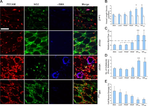
Figure 7.
Coordinated expression of VEGF and PDGF-BB reverses hindlimb ischemia. Ischemic hindlimbs of SCID mice were treated with vehicle (BSA) or myoblasts expressing only β-galactosidase (LacZ), PDGF-BB, VEGF, or both at coordinated levels (VIPlow and VIPhigh, expressing two different average VEGF levels). Angiogenic response (A, B) and regional blood flow (C) were assessed on the distal thigh muscles (quadriceps femoris and biceps femoris), where cells were injected. Collateral growth (D) was assessed in the proximal adductor muscle, which is anatomically remote from the injection sites, and muscle damage (E) was determined in the calf muscles (tibialis anterior and gastrocnemius), where ischemia was maximal. A) Immunostaining of vessels in implanted muscles with antibodies against endothelial CD31 (PECAM; red), pericyte NG2 (green), or smooth muscle αSMA (blue). Scale bar = 50 μm; n = 5–7. B) VLD quantification (micrometers of vessel length per muscle fiber in the area of effect, n=5–7) in the treated muscles (blue bars) and contralateral nonischemic muscles (white bars). C) Relative blood flow in ischemic muscles (percentage of the contralateral nonischemic leg, n=5–7). Dotted line represents normal flow (100%). D) Number of collaterals in the proximal adductor muscle group of treated legs (n=3). E) Damaged tissue in ischemic calf muscles of treated limbs (percentage area of the total muscle on histological sections, n=5–6). *P < 0.05; **P < 0.01.

