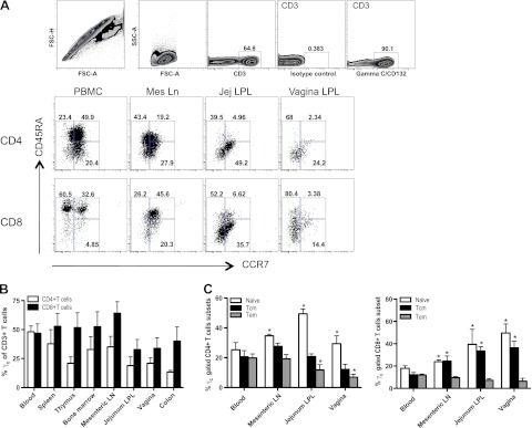
Figure 1.
Surface expression of γc on T cells in various tissues of normal macaques. A) Representative dot plots showing the gating strategy for identifying γc+ CD3+ T lymphocytes and distribution of γc+ CD4+/CD8+ T-cell subsets, defined here as naïve, CD45RA+CCR7+; effector memory, CD45RA+/CD45RA−CCR7−; and central memory, CD45RA−CCR7+, in different tissues. Mes Ln, mesenteric lymph node; Jej LPL, jejunum lamina propria lymphocyte. B) Frequency of γc+ CD4+ (open bars) or γc+CD8+ (solid bars) as a percentage of gated CD3+ T cells in peripheral blood and various lymphoid tissues from normal Rhesus macaques. C) Group data showing the mean frequency of naive (CD45RA+CCR7+), central memory (Tcm; CD45RA-CCR7+), and effector memory (Tem; CD45RA+/CD45RA−CCR7−) gated γc+ CD4/CD8+ T cells in blood, mesenteric lymph node (LN), jejunum lamina propria lymphocyte (LPL), and vagina (n=7 for each tissue). Error bars represent means ± se. *P < 0.05 vs. peripheral blood.

