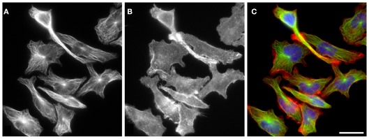
Figure 2.
Distribution of microtubules and F-actin in RBL-2H3 cells. Microtubules (A) and F-actin (B) have distinct subcellular organization in resting interphase RBL-2H3 cells. While microtubule arrays radiate from perinuclear centrosomes, F-actin does not have a single organizing center within the cell and is more concentrated at cell periphery. Cells were fixed with formaldehyde and extracted with Triton X-100 before staining with rabbit antibody to α-tubulin [(A); green] and rhodamine-conjugated phalloidin [(B); red]. DNA was stained with DAPI (blue). Superposition of α-tubulin and F-actin is shown in (C). Scale bar, 20 μm. Photography E. Dráberová (Institute of Molecular Genetics AS CR, Prague).

