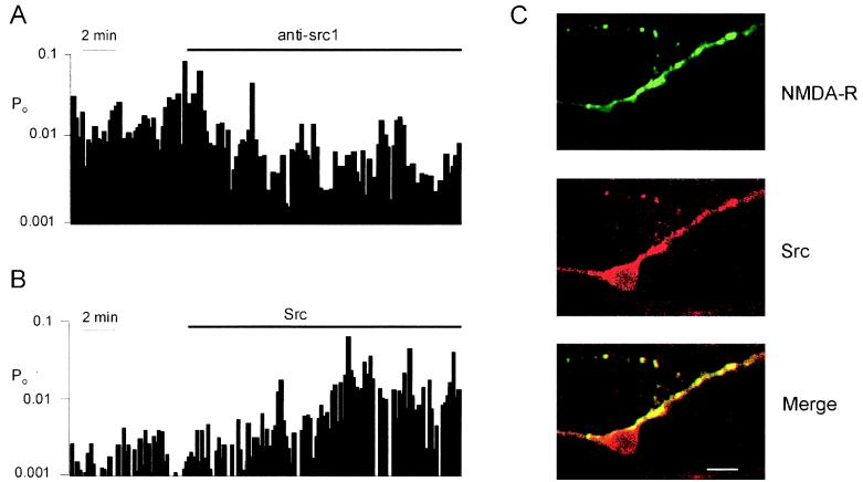
Figure 2.
Regulation of NMDA receptor single-channel activity in inside-out patches by Src and overlapping distribution of Src and NMDA receptor subunit proteins. (A)A continuous record of NMDA channel-open probability (Po). Anti-src1 was applied to the cytoplasmic face of the patch during the period indicated. Po was calculated in bins of 10 sec. (B) A record of NMDA channel Po from a different inside-out patch with Src applied to the cytoplasmic side as indicated. (C) Confocal images show immunofluorescent labeling of a dorsal horn primary culture by antibodies recognizing NR2A/B subunit proteins (green; courtesy of R. Wenthold, National Institutes of Health, Bethesda, MD) or Src (red). Bottom shows the merged images; areas showing overlapping fluorescence are yellow. We found similar colocalization when anti-NR1 and anti-Src antibodies were used. Also, experiments without primary antibodies or with primary antibodies incubated with the respective immunogen peptides showed no labeling. (Bar = 10 μm.)

