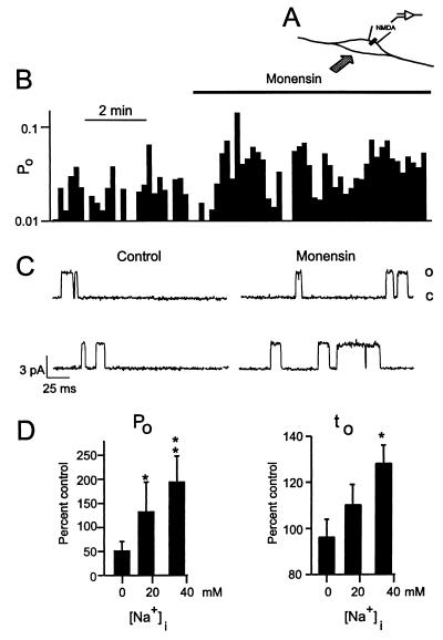
Figure 3.
Increases in [Na+]i by application of monensin potentiate single NMDA channel activity recorded in the cell-attached configuration. A shows the recording configuration. (B) A continuous record of NMDA channel Po from neurons bathed with extracellular solution containing 50 mM Na+. (C) shows representative single-channel currents before and during monensin application. D Changes in Po and mean open time (to) versus [Na+]i during monensin application. For each Na+ concentration, six patches were tested. ∗, P < 0.05; ∗∗, P < 0.01, Mann–Whitney U test when compared with the channel activity at 0 mM Na+.

