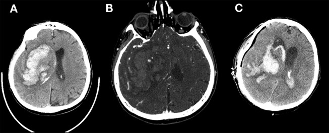
Figure 1.
A 61-year old male patient presenting within 80 min of symptom onset. (A) Axial non-contrast CT demonstrates massive intraparenchymal hemorrhage projected over the basal ganglia with severe mass effect and ipsilateral ventricular hemorrhage. (B) Multiple Spot Signs were present on CTA raw images. (C) Patient underwent emergent craniectomy and hematoma evacuation demonstrated 1 day later.

