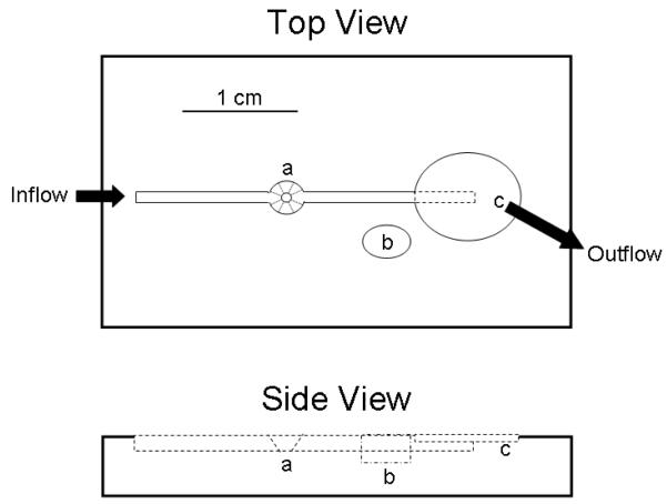
Figure 1. Diagram of a simple flow-cell design for oocyte electrophysiology.
The inflow channel is sized to accommodate small PTFE tubing (OD 0.12″). The oocyte chamber (a) is beveled to allow microelectrodes to enter at an angle from either side of the flow channel. The guard chamber (b) enables use of a salt bridge to reduce grounding variability. The outflow incorporates a stepped chamber design that allows suction to remove excess superfusate without removing all the fluid in the flow channel if inflow stops.

