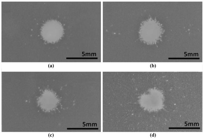
Fig. 7.
Transversal lesion patterns: (a) in a free field, (b) with the rib phantom positioned at 8 cm from the focus, (c) with the rib phantom at 4 cm, and (d) with porcine ribs at 8 cm. Lesions correspond to the visually clear areas surrounded by the darker background color of the red blood cell layer. Collateral damage was defined as the sum of all damage spots detected outside the continuous portion of the main lesion.

