Figure 1.
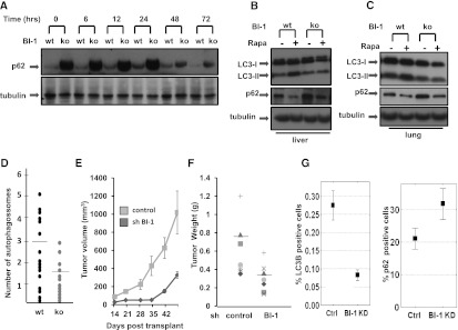
BI-1-deficient mice have reduced autophagy. (A) Protein levels of p62 were assessed by immunoblot analysis of hearts from wild-type (wt) and BI-1 knockout (ko) mice treated with rapamycin for various times as indicated (in hours). Lysates were normalized for total protein content and analyzed by SDS-PAGE/immunoblotting using anti-p62 or anti-tubulin antibodies. Wild-type (wt) and BI-1 knockout (ko) livers (B) and lungs (C) were collected after 24 h of injection with rapamycin. Tissues lysates were analyzed by immunoblotting as above using anti-LC3, anti-p62, and anti-tubulin antibodies. (D) Wild-type (wt) and BI-1 knockout (ko) mice were injected with rapamycin. After 24 h, hearts were dissected, processed, and examined by transmission electron microscopy. From 20 images each for wild-type (wt) and knockout specimens, the total number of autophagosomes was determined using ImageJ software. Mice were injected with 1 mg of rapamycin per kilogram of body weight. Horizontal bars indicate mean. Data are statistically significant by t-test (P = 0.009). (E) Female BALB/c nu/nu mice were injected subcutaneously with H322M cells (5 × 10⁶) containing scrambled control or BI-1 shRNA vectors. Tumor volumes were measured over time (mean ± SD; n = 10). (F) Seven weeks post-transplantation, animals were sacrificed, and tumors were excised and weighed (mean ± SD; n = 10 animals per group). (G) Tumor sections were analyzed for LC3 and p62 staining by quantitative immunohistochemistry. The overall low percentage of cells showing punctate LC3 immunostaining may reflect the antibody conditions employed, which were designed to avoid detection of diffuse cytosolic staining of nonautophagic cells.

