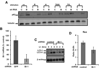
Figure 2.
BI-1 knockdown reduces autophagic flux. (A) Stably transduced H322M cells were cultured in standard rich medium or with serum- and glucose-deficient medium ([H] HBSS; [E] EBSS) alone or with various agents, including rapamycin (R; 25 μg/mL), thapsigargin (TG; 5 μM), and tamoxifen (TAM; 10 μM), for 16 h. Lysates were prepared, normalized for total protein content, and analyzed by SDS-PAGE/immunoblotting using anti-p62 and anti-tubulin antibodies. (B) H322M cells were stably transduced with recombinant shRNA lentiviruses targeting BI-1 or GFP. Relative levels of endogenous BI-1 mRNA were assessed by quantitative RT–PCR (expressing data as a percentage of control relative to cells transduced with GPF shRNA control vector). (C) H322M cells that were stably transduced with recombinant shRNA lentiviruses targeting BI-1 or GFP (control) were treated with LIs (20 mM NH4Cl and 100 μM leupeptin) for either 2 h or 4 h, as indicated. Levels of LC3-I and LC3-II were analyzed by immunoblotting. Blots were reprobed with anti-β-actin antibody as a loading control. (D) Bands were quantified by densitometry, and measurements were used to calculate LC3 flux (mean ± SD; n = 3 experiments; P = 0.017).

