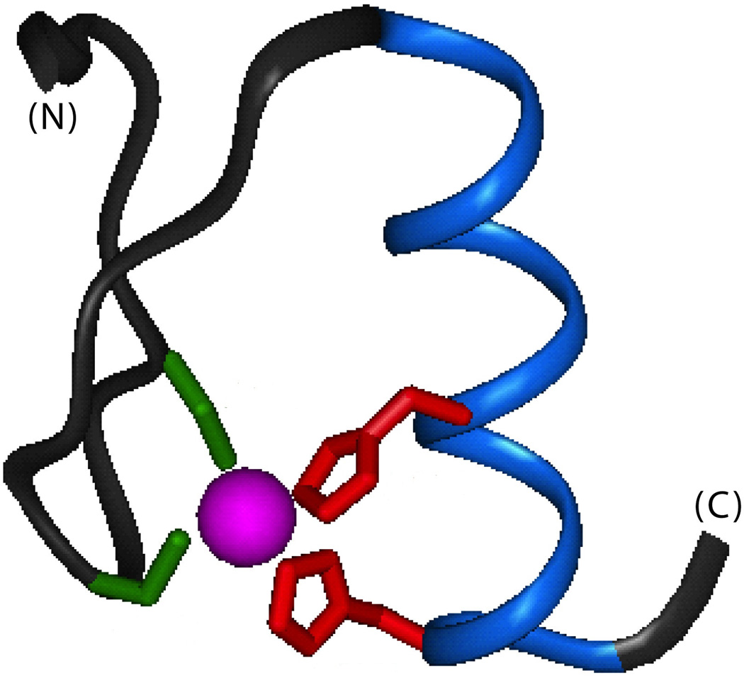
Figure 7.
Classical zinc-finger motif. The peptide backbone is shown in black (N-terminal β-hairpin and central loop) or blue (C-terminal α-helix); the encaged zinc ion is shown in magenta. The interior Zn2+ coordination site of the C2H2 motif comprises the thiolate groups of two cysteine side chains (green) and imidazole rings of two histidine side chains (red). The structure shown is domain 2 of Zif268;85 coordinates were obtained from Protein Databank entry 1ZAA.

