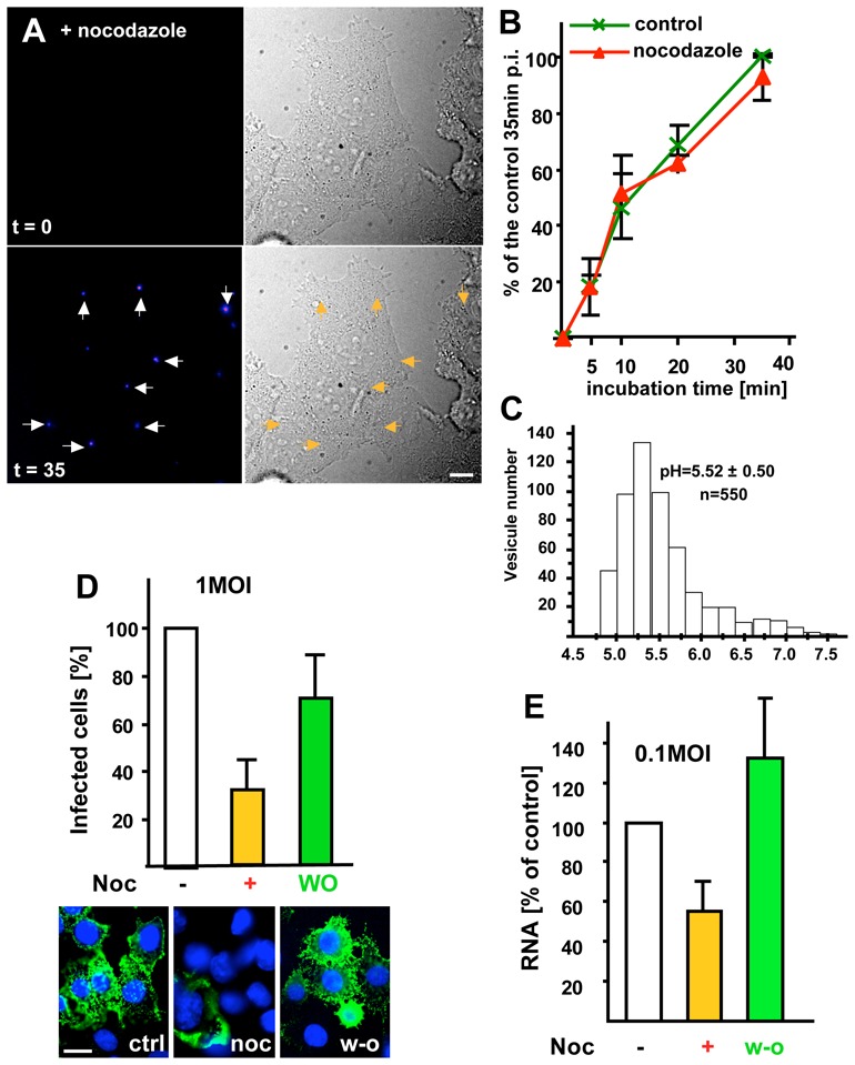
Figure 2. Microtubule-dependent transport.

(A) Viral fusion was studied as in Fig 1A in cells pre-treated with 10μM nocodazole for 2h (the drug was present throughout the experiment), and frames were captured at the indicated time. (B) After treatment with or without nocodazole as in (A), the number of cells containing fused viruses was counted at the indicated times during the 37°C incubation. Values are expressed as a percentage of the untreated control after 35min, as in Fig 1B. (C) After microtubule depolymerization as in (A), BCECF-dextran was endocytosed for 10 min at 37°C and chased for 35min. The pH of individual endosomes was measured by BCECF-dextran fluorescence ratio imaging. The histogram shows the pH distribution of 550 endosomes with the means ± SD indicated on the figure. (D) VSV (1MOI) was bound at 4°C to the surface of BHK cells preincubated without (control, ctrl) or with nocodazole (noc), as in (A). Cells were incubated for 3h at 37°C to allow infection to proceed. When indicated (W-O, wash out), nocodazole was removed, and incubation continued without drugs for 2h. Cells were analyzed by immunofluorescence microscopy using antibodies against VSV-G after labeling nuclei with DAPI (blue). Typically, ≈70% of the cells were infected under control conditions, and the number of infected cells is expressed as a percentage of the untreated control. (E) Cells treated or not with nocodazole (as in D) were infected with 0.1 MOI VSV. Replication of VSV RNA minus strand was quantified by TaqMan-RT-PCR and results are expressed as a percentage of the untreated control. Number of experiments: B, 3; D, 6; E, 4. Bars, A: 2,5μm; D: 4μm.
