
Figure 8. Nucleocapsid release in vitro.
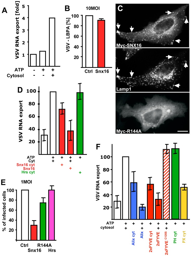
(A) Late endosomes were loaded with VSV in vivo, and endosomal fractions were prepared. These fractions [18μg] were incubated in vitro with an ATP-regenerating (ATP), with or without cytosol for 20 min at 37°C. Then, free (cytosolic) RNA was separated from endosome-associated RNA by floatation in a sucrose gradient, and VSV RNA minus strand was quantified by TaqMan-RT-PCR. RNA export is expressed as the ratio of free RNA released in the presence of ATP and cytosol over the negative controls without cytosol and ATP. (B) VSV endocytosed for 45 min (as in Fig 1E) in cells expressing Snx16-myc was analyzed by fluorescence microscopy; VSV-G colocalization with LBPA was quantified as in Fig 7D. (C) After expression of WT Snx16-myc or Snx16R144A-myc, HeLa cells were processed for immunofluorescence using antibodies against myc and Lamp1 (upper panels, double immuno-fluorescence) or myc alone (lower panel). (D) The assay was as (A) with cytosol prepared from cells overexpressing Snx16 (Snx16 cyt) or Hrs (Hrs cyt), or with control cytosol supplemented with 0.5μg purified recombinant Snx16 (Snx16). (E) Cells expressing or not Snx16, Snx16R144A or Hrs were infected with VSV (1MOI), and analyzed by fluorescence microscopy; infected and non-infected cells were quantified as in Fig 2D. (F) The assay was as in (A) with cytosol prepared from cells overexpressing GFP-2xFYVE (2xFYVE cyt), Alix (Alix cyt), the GFP-PH domain of PLC™ (PH cyt) or the GFP-PX domain of p40phox (PX cyt). Alternatively, the assay was carried out with control cytosol supplemented with 0.5μg purified, recombinant Alix, GST-2xFYVE (2xFYVE) or GST-2xFYVEC125S (2xFYVEC125S). VSV RNA− export was expressed as a percentage of the positive control, to facilitate comparison between different experiments. Number of experiments: A, 6; B–E 3; F, 4. Bar, C: 2,5μm.