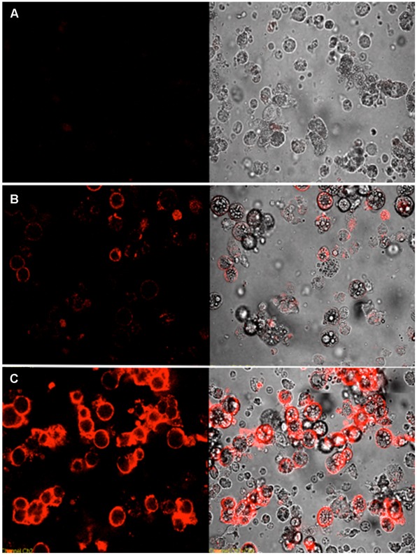
Figure 3. Confocal images of aptamers stained with cultured differentiated 3T3-L1 adipocytes.
Cells were incubated with aptamers conjugated with biotin, and binding events were observed with PE-conjugated streptavidin. (A) unselected PE-labeled library; (B) adipo-1; (C) and adipo-8. The final concentration of the aptamers in the binding buffer was 250 nM. (Left) fluorescence and (Right) optical images of differentiated 3T3-L1 adipocytes.

