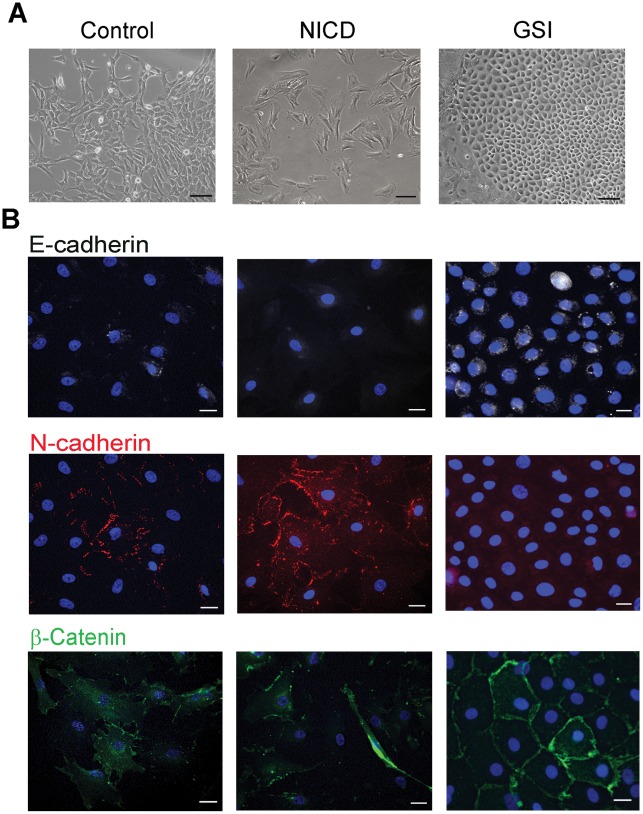
Figure 5. Suppression of Notch reversed mesenchymal phenotype of explant-derived cells.
C-Kit+ and c-Kit- cells were cultured in presence of GSI. Representative images of c-Kit- cells are shown. (A) Transmitted light images demonstrate changes in cell morphology upon treatment with NICD or GSI. (B) Cells were labeled with antibodies to E-cadherin (white), N-cadherin (red) or β-catenin (green) as indicated. Nuclei were counterstained with DAPI (blue). Scale bars, 100 µm (A) or 20 µm (B).

