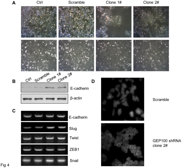
Figure 4. GEP100 down-regulation induces epithelial change of cancer cells and up-regulation of E-cadherin protein.
(A) A mesenchymal to epithelial change was observed in the cells stably knocked-down for GEP100. The upper and lower panels showed the morphological appearances at a low cell density and when the cells reached confluence, respectively. Comparing with the control and scramble groups, the cell in the experimental group became epithelial-like and adhesive. (B) Western blot showed that the expression level of E-cadherin protein was increased about 3-fold in the experimental group compared with the control group. (C) RT-PCR analysis of E-cadherin mRNA and its transcription regulators. The expression of E-cadherin mRNA was not affected by GEP100 down-regulation. An increase of Slug mRNA was found in the GEP100 knocked-down cells, while there was no change for Twist, ZEB1 and Snail. (D) Immunofluorescence staining of E-cadherin. Comparing with the scramble group, GEP100 down-regulation redistributed the E-cadherin into the cell-cell contacts.

