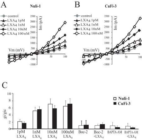
Figure 4. Dose dependency of the effect of LXA4 on whole-cell currents of normal (Nuli-1) and CF (CuFi-3) bronchial epithelial cell lines.
Typical I-V relationships obtained before and after 10 min exposure to 1 pM (n = 6 Nuli-1, n = 6 CuFi-3), 1 nM (n = 4 Nuli-1, n = 4 CuFi-3), 10 nM (n = 3 Nuli-1, n = 3 CuFi-3) and 100 nM (n = 6 Nuli-1, n = 6 CuFi-3) LXA4 in Nuli-1 (A) and CuFi-3 (B) cell lines. (C) Mean inward conductance changes normalized to control values (gi/gic obtained without LXA4) as a function of LXA4 concentration in Nuli-1 (open bars) and CuFi-3 (black bars) cells in control conditions and obtained upon exposure to Boc-2 (10 µM) alone (10 min, 100 nM, n = 4 Nuli-1, n = 4 CuFi-3) or with Boc-2 (10 µM) and LXA4 (10 min, 100 nM, n = 6 Nuli-1, n = 6 CuFi-3) and after BAPTA-AM pre-treatment alone (n = 6 Nuli-1, n = 6 CuFi-3) or with BAPTA-AM and LXA4 (10 min, 100 nM, n = 4 Nuli-1, n = 6 CuFi-3).

