
Figure 6. Deviation from independence, as a function of tissue type.

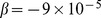
The overall deviation of the joint distribution from the expected independent distribution was scored using JSD for PC (A) and PE (B). The lowest JSD was observed in lung PE ( ), while the highest was observed in liver PE (
 ). During heart development from day
  to day
  , a trend of decreasing JSD was observed in PC (
 ,
,  ), while PE displayed stable JSD during the time period, (
 ,
,  ).
).