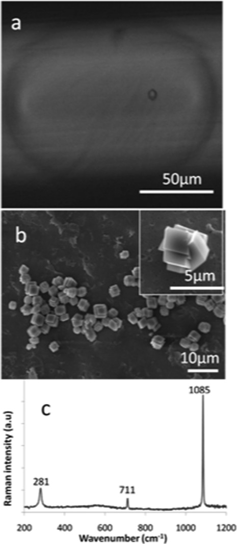
Figure 4.
(a) Photomicrograph of an individual droplet formed in the microfluidic device approximately10 s after the start of the reaction. The droplet contents were formed by combination of 4 mM CaCl2 and Na2CO3 solutions, and a single rhombohedral calcite particle can clearly be seen within the droplet. (b) SEM micrograph of CaCO3 particles isolated from microdroplets formed by combination of 4 mM CaCl2 and Na2CO3 solutions. Only rhombohedral crystals between 1 and 2 μm in size were obtained. The inset shows an individual particle at higher magnification. (c) Raman spectrum of one of the crystals shown in (b).

