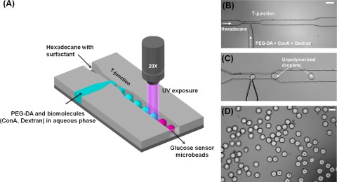
Figure 2.
(A) Schematic for the generation of PEG based glucose sensing microbeads showing two inlets at the T-junction to form droplets encapsulating PEG-DA and biomolecules in an aqueous phase. The droplets were photopolymerized by UV light illuminated from a 20× microscope lens to form PEG microbeads. (B) Photomicrograph of T-junction exhibiting two inlets for droplet generation (Scale bar = 200 µm). (C) Generation of unpolymerized PEG-DA droplets at the T-junction. (D) Photopolymerized PEG-DA microbeads collected outside the microdevice (Scale bar = 100 µm).

