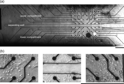
Figure 3.
(a) 5× differential interference contrast (DIC) image of a neuronal network grown within the microfluidic device. In the middle, it is possible to observe the 100 μm wall which creates two symmetric compartments without affecting the electrode functionality. The image is a combination of 16 individually 5× pictures. (b) High resolution images of neurons cultured within the device in the (I) lower and (III) upper compartment, and (II) in the region across the separating wall. Scale bars are 500 μm.

