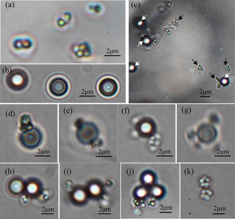
Figure 5.
Attachment of PS microbeads to the MO-1 cells under a phase-contrast microscope. (a) MO-1 cell, (b) PS microbeads, (c) MO-1 cells attached with antibody-coated microbeads, the black arrows indicate cells and the white arrows indicate PS microbeads, (d) one microbead and one cell, (e) one microbead and two cells, (f) one microbead and three cells, (g) one microbead and four cells, (h) two microbeads and two cells, (i) two microbeads and three cells, (j) three microbeads and two cells, and (k) two cells.

