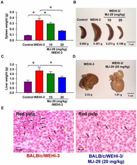
Figure 7. Effects of the weights in spleen and liver as well as histopathological examination of spleen tissues on MJ-29-treated leukemic mice.
Animals were intravenously injected with WEHI-3 cells (1×10⁶ cells/100 µl per mouse) in PBS, and then treated intraperitoneally with MJ-29 (10 and 20 mg/kg alternate day for 8 times). Weights and representative images of spleen (A and B) and liver (C and D) tissues from leukemic mice were determined and measured individually. Each point is mean ± S.D. (at least five samples). * p<0.05 indicates significant difference by Tukey's HSD test between the WEHI-3 leukemic mice and experimental (normal or intraperitoneal treatment with MJ-29 at 10 and 20 mg/kg, respectively) groups. (E) Dissected leukemic mice and hematoxylin-eosin stain for the paraffin sections of spleens from MJ-29-treated and un-treated leukemic mice as described in the “Materials and Methods”. Arrows (↑) shows infiltration of immature myeloblastic cells (leukemia cells) into red pulp of the spleen. R, red pulp. The data are performed with representative experiment in triplicate and three independent experiments with similar results.

