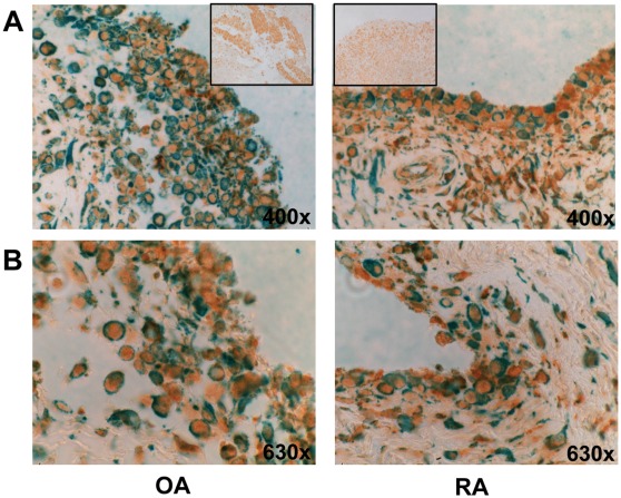
Figure 3. Immunohistochemistry.
Representative photomicrographs showing immunohistochemical double staining of OA and RA synovial tissues with P2X4 (brown) and fibroblast marker (green). A. original magnification 400×, box shows the Isotype control, magnification 200×. B. original magnification 630×.

