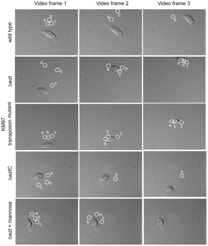
Figure 1. Image frames from various time points of live-cell microscopy videos of rainbow trout skin epithelial cells following infection with various V. anguillarum strains.
Using live-cell microscopy, a video clip (Video S1, S2, S3, S6, and S7 supporting information) was recorded of the phagocytic activity of rainbow trout skin epithelial cells after infection with the wild type, the KM97 transposon mutant, the Δwzt mutant, and the Δwzt mutant complemented with the wild-type wzt gene. Three frames from each video progressing in time from left to right are shown indicating the phagocytic activity of an epithelial cell after contact with each strain. Similar videos were made for the Δwzm and ΔwbhA mutants, and similar results to that of the Δwzt mutant were seen (supporting information Videos S4 and S5). Thus, the Δwzt mutant results are given as a representative for all three mutant strains. In the first frame from each video, three bacterial cells were circled and labeled. These three bacteria were followed in the two additional time frames to indicate movement of the bacteria during the movie. If the cell is no longer labeled in frames 2 and 3, then the bacterial cell was displaced from the glass surface and swam away. In the bottom set of frames, the epithelial cells were pretreated with 1 mM mannose before infection with the Δwzt mutant. The three-image sets are designated by the strain mutation to the left and mutation designations followed by a C are complemented.

