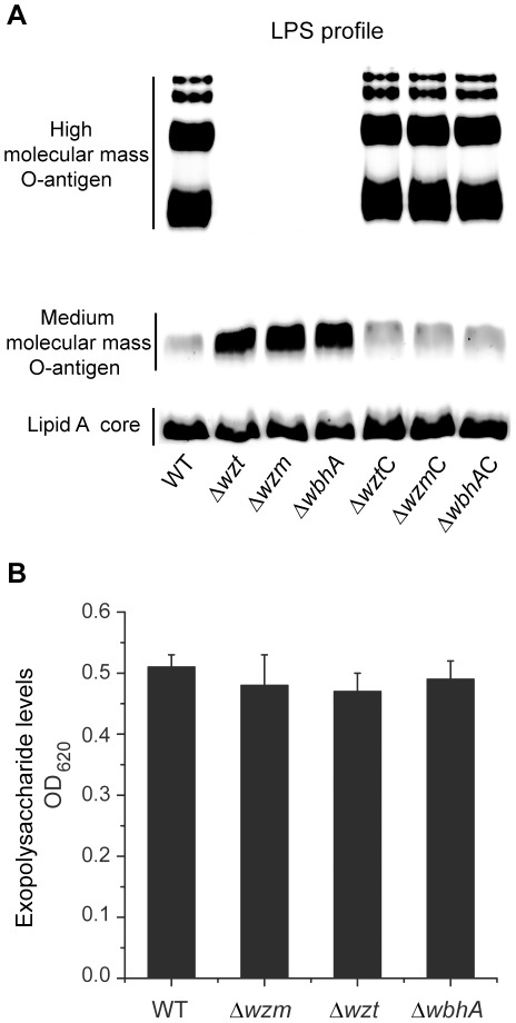
Figure 3. Lipopolysaccharide and exopolysaccharide analyses.
(A) Lipopolysaccharides were fractionated by 15%-SDS-PAGE and detected using the Pro-Q® Emerald 300 Lipopolysaccharide Gel Stain Kit (Invitrogen). (B) Exopolysaccharides were extracted from culture supernatants and quantified using an Alcian blue technique described by Plante [56]. The amount of EPS is given as an OD620 reading. For both (A) and (B), strains are designated by their mutation. Mutation designations followed by a C are complemented with the respective wild-type gene.

