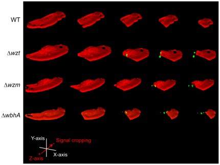
Figure 4. Confocal microscopy 3D-imaging of rainbow trout skin epithelial cells following infections.
Skin epithelial cells were infected with the wild type and the Δwzt, Δwzm, and ΔwbhA mutants. Following fixation, the epithelial cells were visualized by labeling actin with Alexa Fluor 568 phalloidin (red). After permeabilization of the epithelial cell, bacteria were labeled using a whole-cell antiserum and a FITC-conjugated Donkey anti-rabbit IgG (green). Using the NIS-elements AR 3.2 software, a 3D rendering of a confocal microscopy z-stack covering an entire cell was done and is presented as the left most image for each strain. In each image, the phalloidin (red) signal was progressively cropped along the z-axis to reveal internal bacteria. Strains are designated by their mutations and the designations are given to the left of the image.

