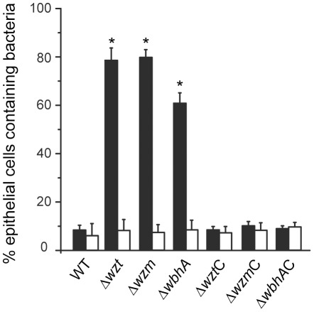
Figure 5. Quantification of phagocytic activity by rainbow trout skin epithelial cells following infection with various V. anguillarum strains.
Epithelial cells were isolated from the skin of rainbow trout and either left untreated (closed bars) or pretreated with 1 mM mannose (open bars) before infection with 105 bacteria ml−1. Following a 3 h infection, the epithelial cells were fixed and the epithelial cells and bacteria were fluorescently labeled. Confocal 3D-imaging microscopy was done on at least 100 single epithelial cells from three separate experiments to determine the number of cells that contained bacteria. These data are presented as a percent. The V. anguillarum strains that were tested are designated by their mutation. Mutation designations followed by a C are complemented with the respective wild-type gene. Asterisks indicate a p-value of <0.05 as determined by the Student's t-test.

