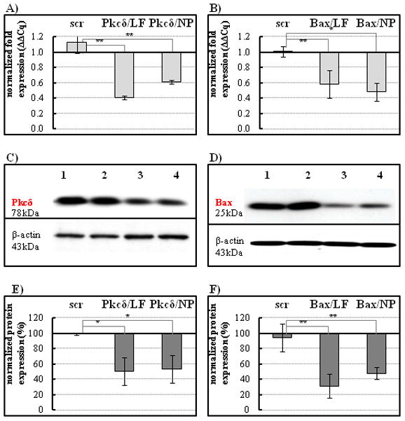
Figure 2.

siRNA knock down of Pkcδ or Bax expression in cultured salivary gland cells. A–B, mRNA expression was analyzed by qPCR 24 hours following transfection. A, Transfection with Pkcδ siRNA using Lipofectamine (LF) or nanoparticles (NP) inhibits Pkcδ mRNA expression compared to scrambled siRNA control (scr) delivered by nanoparticles; B, Transfection with Bax siRNA using Lipofectamine (LF) or nanoparticles (NP) demonstrated efficient reduction of Bax mRNA expression levels. Results are representative of three replicates and compared to untreated controls (fold changes); *P < 0.05 by one-way analysis of variance with Dunnett’s multiple comparison test. C–D, The lysed cells were analyzed for expression levels of the two pro-apoptotic proteins on Western blots. C, Pkcδ, or D, Bax protein expression was normalized to β-actin (blots were stripped and reprobed). Lane 1, untreated cells; lane 2, cells treated with scrambled siRNA control; lane 3, cells treated with Lipofectamine-mediated siRNA, as positive control; lane 4, cells treated with nanoparticle-mediated siRNA. E–F, graphs show quantification of protein band intensities from Western blot analysis, scanned by alpha imaging and measured using the ImageJ (NIH) software. LF, Lipofectamine; NP, nanoparticle complexes. Calculations were based on three measurements compared to untreated controls (protein level established as 100%); *P < 0.05, **P< 0.001.
