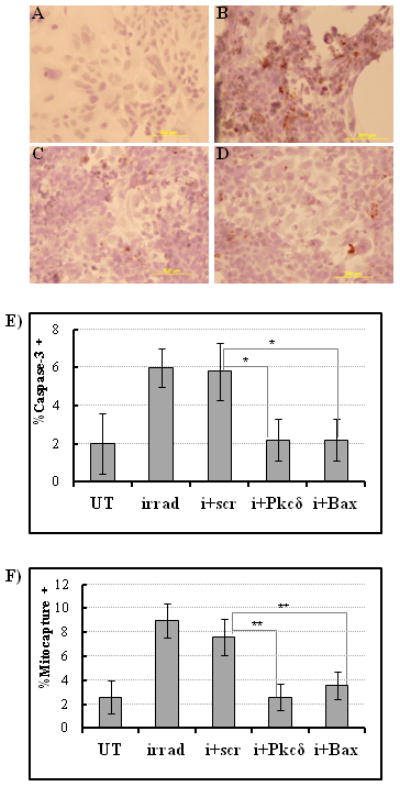
Figure 4.

Inhibition of pro-apoptotic Pkcδ or Bax protein expression decreases salivary gland cell sensitivity to radiation damage in vitro. A, Non-irradiated control cells stained with caspase-3 antibody and hematoxylin show no caspase-3 activity. B, Radiation treatment alone significantly increases caspase-3 activity in salivary gland monolayer cultures (caspase-3 positive cells are dark brown on hematoxylin counterstain). Pre-treatment with (C) nanoparticle/Pkcδ siRNA complexes or (D) nanoparticle/Bax siRNA complexes reduces caspase-3 staining in irradiated salivary gland cells compared to non-treated cells. E, Quantitative analysis of the caspase-3 staining based on cell counts within a defined region of the microscopic field. Percentage of caspase-3 positive cells is derived from number of caspase-3 positive cells/ total number of cells counted. UT, untreated control cells; irrad, irradiated cells; i+scr, irradiated cells pretreated with scrambled siRNA; i + Pkcδ or Bax, irradiated cells pretreated with siRNA to Pkcδ or Bax (n=5, *P < 0.05). F, Mitochondrial membrane disruption was determined using a Mitocapture assay 24 hours after irradiation (n=5 randomly chosen microscopic fields of each sample group) Percentage of mitocapture-positive cells is derived from # of mitocapture positive cells/ total # cells counted. UT, untreated cells; irrad, irradiated cells; i+scr, irradiated scrambled-siRNA treated cells; i+ Pkcδ, irradiated Pkcδ-siRNA treated cells; i+Bax, irradiated Bax-siRNA treated cells. **P<0.001. Statistical significance was tested by ANOVA and Tukey’s multiple comparison tests.
