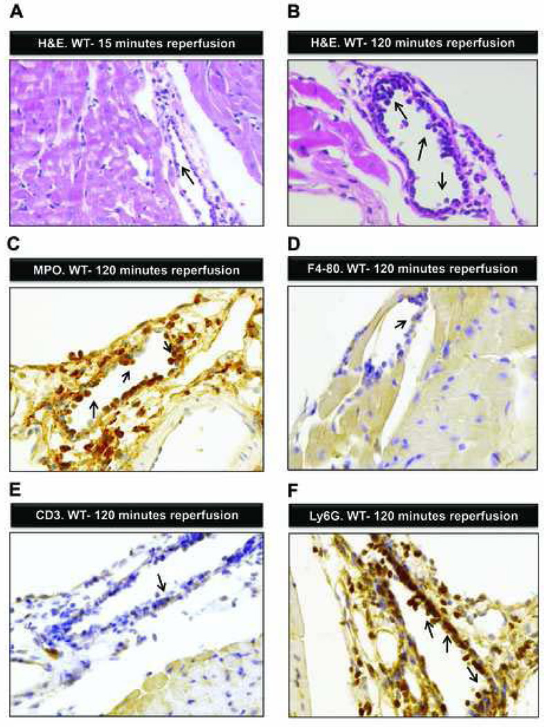
Figure 4. Ischemia-reperfusion associated histological changes.
(A) Hematoxylin and eosin stain staining of the myocardial tissue after 60 min of ischemia and 15-min reperfusion. The cardiac structure appears normal with a few inflammatory cells (arrow) adhering to the endothelia. (B) Hematoxylin and eosin staining of the myocardium after 60 min of ischemia followed by 120 min of reperfusion. Multiple inflammatory cells adhering to or passing the endothelial cell layer (arrows; original magnification 400×). (C) Immunohistochemistry for Myeloperoxidase of myocardial tissue after 60 min of ischemia and 120 min of reperfusion. Multiple Myeloperoxidase positive cells adhere to the endothelial cell layer or transmigrate to the perivascular space (arrows; original magnification 200×). (D) Immunohistochemistry for F4–80 surface protein (expressed on monocytes/macrophages) after 60 min of ischemia by 120 min of reperfusion. Single F4–80+ cell adheres to the endothelial cell layer (arrow). (E) Immunohistochemistry for CD3 (T-cell-marker) after 60 min of ischemia and 120 min of reperfusion. Sporadic CD3+ cells attach to the endothelial cell layer (arrow). (F) Immunohistochemistry for Ly6G (polymorphonuclear leukozyte (PMN) marker) after 60 min of ischemia and 120 min of reperfusion. Multiple PMNs adhere to the endothelial cell layer and infiltrate the myocardium (arrows, original magnification 200×); shown are representative images from one experiment out of four mice.

