
Figure 3.
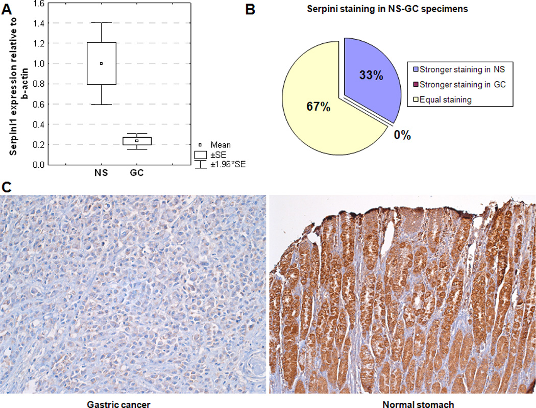
Serpini1 is expressed at lower levels in gastric cancer (GC) vs. normal stomach (NS).
Panel A. mRNA levels of Serpini1 are lower in GC than in NS. X-axis: specimens; Y-axis: mRNA level of Serpini1 relative to beta-actin.
Panel B. Approximately 2/3 of specimens demonstrated similar Serpini1 staining in GC and matched NS (green in the figure). Approximately 1/3 of specimens demonstrated stronger staining in NS than in matched GC (blue in the figure)
Panel C. Representative image of staining with Serpini1 antibody in GC and NS, demonstrating stronger staining in NS than in GC.