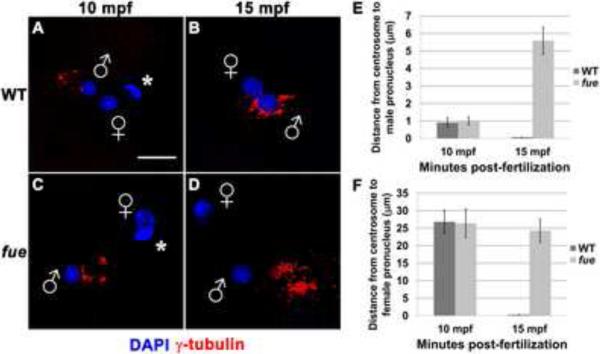
Figure 1. Centrosomes fail to attach to pronuclear envelopes in fue embryos.
In vitro fertilized embryos from wild-type and fue females fixed and labeled for centrosomes (γ-tubulin antibody, red) and DNA (DAPI, blue) at 10 (A,C) and 15 mpf (B,D). Asterisks indicate polar bodies, and male and female pronuclei are indicated with symbols. Scale bar represents 20 μm and applies to all panels. Images are projections from confocal z-stacks. (E,F) Distance between centrosomal γ-tubulin labeling and male or female pronuclear envelopes quantified at 10 and 15 mpf. Error bars indicate +/- 1 standard error. See also Figure S1, and Movies S1 and S2.

