
Fig. 3.
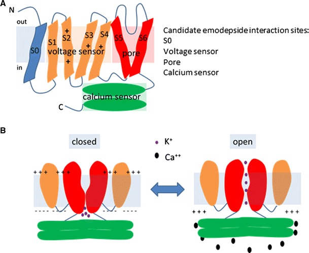
A schematic of the structure of the SLO-1/BK calcium-activated potassium channel. This is based on the information from papers that have reported the structural properties of the human BK channel (Latorre et al. 2010; Pantazis et al. 2010; Yuan et al. 2010, 2012) and a review of these data (Latorre et al. 2010). a A single α subunit of the SLO-1 channel showing the relationship between the voltage sensor domain, pore forming domain and calcium sensing domain. S0 is the least conserved region between the different isoforms of the channel, whereas the pore forming and calcium sensing domains are highly conserved. The transmembrane segments S2, S3 and S4 harbour charged residues as indicated which confer the voltage sensitivity of the channel. b A simplified diagram (omitting S0) showing the relationship between two α subunits of the SLO-1 tetramer and the opening of the channel