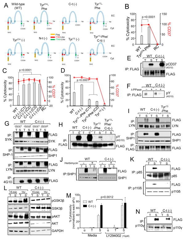
Figure 4. CD37 possesses dual inhibitory and activation signaling function.
(A) Schematic representation of the human CD37 protein showing the mutations introduced in the cytosolic regions. (B–D) CD37+FLAG 697 WT or mutant cell lines were treated with SMIP-016+αFc. Cell death was assessed at 24 hr by annexin-V/PI assay (gray bars). Data shown were normalized to αFc alone (B and C, n=10; D, n=16). Expression of CD37 was assessed by flow cytometry (red lines). RC= Cells expressing a reverse complement sequence of CD37. (E) Lysates from trastuzumab+αFc (T) or SMIP-016+αFc (S) treated WT or C-t(−) cells (15 min) were immunoprecipitated with anti-FLAG followed by 4G10 or FLAG immunoblot. (F) A portion of the immunoprecipitated lysates from E was treated with λ-protein phosphatase (λ-PPase) and analyzed by 4G10 immunoblot. Irrelevant lanes were cropped out the gel. (G) Lysates derived from WT or C-t(−) cells treated with trastuzumab+αFc (T) or SMIP-016+αFc (S) (15 min) were immunoprecipitated using anti-SYK, -LYN, -SHP1 or 4G10 antibody followed by immunoblot analysis with the indicated antibodies. (H) Lysates derived from WT, C-t(−), and Tyr13-Phe cells treated with trastuzumab+αFc (T) or SMIP-016+αFc (S) (15 min) were immunoprecipitated with anti-FLAG antibody followed by immunoblot analysis using 4G10 or anti-FLAG antibodies. (I) Lysates derived from WT, C-t(−), and Tyr13-Phe cells treated with trastuzumab+αFc (T) or SMIP-016+αFc (S) (15 min) were immunoprecipitated using anti-LYN, -SHP1 or 4G10 antibody followed by immunoblot analysis with the indicated antibodies. (J) WT or C-t(−) cells were pretreated with either DMSO or herbimycin (10μM) for 45 min before the addition trastuzumab+αFc (T) or SMIP-016+αFc (S) (15 min). Lysates derived from these cells were immunoprecipitated with anti-SHP1 antibody and analyzed by FLAG and SHP1 immunoblot. (K) Lysates derived from WT and C-t(−) cells treated with trastuzumab+αFc (T) or SMIP-016+αFc (S) (15 min) were immunoprecipitated with anti-p85 or p110δ antibodies followed by immunoblot with the indicated antibodies. Representative of two experiments. (L) Lysates derived from trastuzumab+αFc (T) or SMIP-016+αFc (S) treated WT and C-t(−) cells were analyzed by immunoblot using the indicated antibodies. (M) WT and C-t(−) cells were pretreated with LY294002 for 45 min before the addition of αFc, trastuzumab+αFc or SMIP-016+αFc. Cell death was measured 24 hr post treatment by annexin-V/PI (n=15). (N) Lysates derived from trastuzumab+αFc (T) or SMIP-016+αFc (S) treated WT and C-t(−) cells (15 min) were immunoprecipitated using p110γ antibody followed by FLAG immunoblot (representative of two experiments). Data are represented as mean ± SD for all the relevant panels. See also Figure S2.

