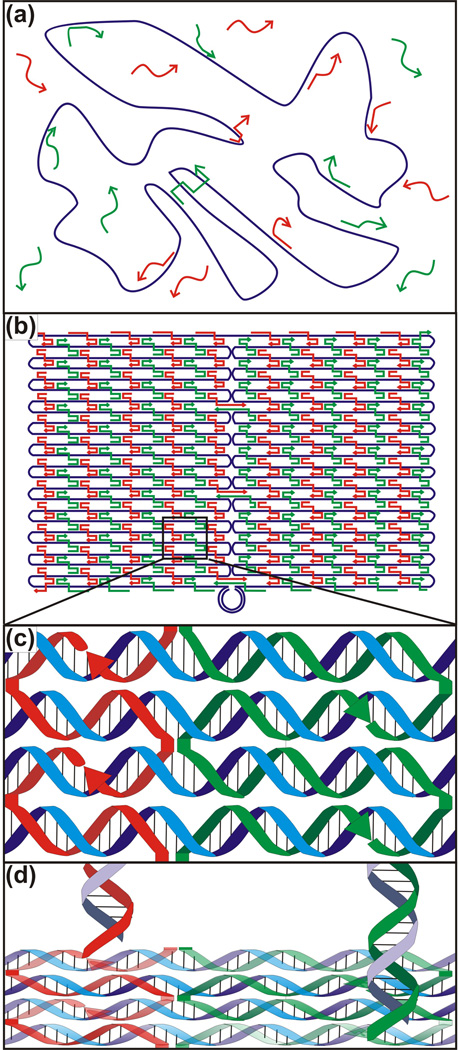
Figure 3.
The DNA origami method. (a) The single-stranded DNA “scaffold” strand (purple) is folded and held in place by specifically hybridizing “staple” strands (red and green). (b) Resultant rectangular origami tile once all the staples have bound to the scaffold. (c) Triple crossover motifs demonstrated by the staples interacting with the scaffold. (d) 5’ ends of the staples are extended to create overhangs, which then can be decorated with partially complementary oligonucleotides (purple).

