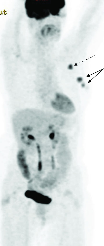
Figure 1.
18F-fluorodeoxyglucose (FDG) positron emission tomography–computed tomography of a patient with newly diagnosed primary breast cancer. The maximum intensity projection image shows multifocal disease in the left breast (solid arrows). Avid left axillary nodal uptake is also identified (dashed arrow). No distant FDG-avid disease is identified.

