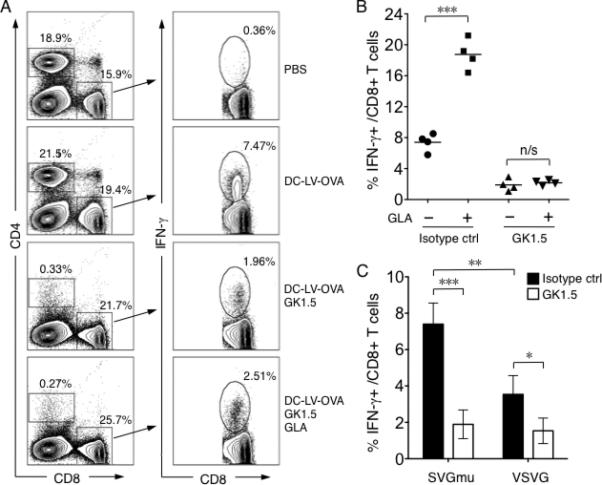
Fig. 5.

The role of CD4+ helper T cells in GLA-enhanced CD8+ T cell responses. A. FACS analysis of intracellular staining of IFN-γ from splenocytes of mice immunized with PBS, DC-LV-OVA, DC-LV-OVA plus GK1.5 antibody-mediated depletion of CD4+ T cells, or DC-LV-OVA plus GLA-SE and GK1.5. One representative data from a group of four mice is shown. Percentage shown in the left panel is the percentage of CD4+ T cells and CD8+ T cells. B. Statistical data showing OVA-specific CD8+ T cells by intracellular staining of IFN-γ following stimulation with OVA257-264 peptide. C. OVA-specific CD8+ T cell percentage by intracellular staining of IFN-γ after OVA257-264 peptide stimulation from mice immunized for 14 d with the same transduction units (TUs) of either SVGmu- (left) or VSVG- (right) enveloped LV-OVA. Black bar: isotype control antibody; white bar: GK1.5 antibody. (***: P < 0.001; **: P < 0.01; *: P < 0.05 and n/s: not statistically significant; One-way ANOVA followed by a Bonferroni's multiple comparison test. Mean +/- SD is shown.)
