
Fig. 7.
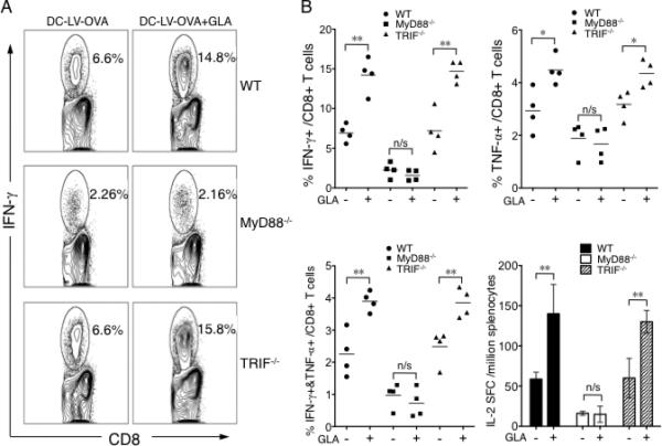
Involvement of MyD88 and TRIF signaling pathways in GLA-adjuvanted DC-LV immunization in vivo. A. Wild-type (upper), MyD88-/- (middle), and TRIF-/- (lower) mice were immunized with DC-LV-OVA, or DC-LV-OVA plus GLA-SE. 2 wk later, OVA-specific CD8+ T cells were analyzed by intracellular staining of IFN-γ following OVA257-264 peptide stimulation. The FACS data shown is representative of four mice tested. B. Statistical data showing OVA-specific CD8+ T cells analyzed by IFN-γ+ (upper left), TNF-α+ (upper right), or IFN-γ+TNF-α+ (lower left) populations for groups of mice described above with (+) or without (-) GLA treatment. (B, lower right) Splenocytes were also pooled for an ELISA assay of IL-2 production after stimulation with OVA323-339 peptide. (**: P < 0.01; *: P < 0.05 and n/s: not statistically significant; One-way ANOVA followed by a Bonferroni's multiple comparison test. Mean +/-SD is shown.)