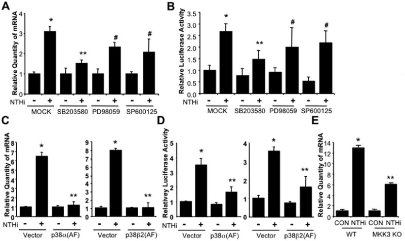
Figure 4. NTHi-induced PAI-1 expression is mediated by MKK3-p38 MAPK.

A & B. PAI-1 induction by NTHi is mediated by p38 MAPK but not ERK or JNK MAPKs in vitro in epithelial cells. Cells were pre-treated with specific chemical inhibitors for p38 (SB203580, 10 μM), ERK (PD98059, 10 μM), or JNK (SP600125, 10 μM) for 1 hour followed by NTHi or saline treatment. PAI-1 mRNA expression was measured by Q-PCR analysis 5 hours after NTHi treatment (A). Cells transfected with PAI-1-Luc were pre-treated with specific chemical inhibitors for p38 (SB203580, 10 μM), ERK (PD98059, 10 μM), or JNK (SP600125, 10 μM) for 1 hour followed by NTHi or saline treatment. PAI-1 transcriptional activity was measured by luciferase assay 5 hours after NTHi treatment (B). C & D. p38 MAPK mediates NTHi-induced PAI-1 expression in vitro in cells. Cells were transfected with control Vector, p38α(AF), or p38β2(AF) for 48 hours and then treated with NTHi or saline. mRNA expression of PAI-1 was measured 5 hours after NTHi treatment by Q-PCR analysis (C). Cells were co-transfected with PAI-1-Luc with or without p38α(AF) or p38β2(AF) for 48 hours and then treated with NTHi or saline. PAI-1 transcription was measured by luciferase assay 5 hours after NTHi treatment (D). E. NTHi induces PAI-1 expression in a MKK3 dependent manner. MKK3 KO and their littermate WT control mice were i.t. inoculated with NTHi, and mRNA expression of PAI-1 was measured from the lung tissues of mice 6 hours after NTHi or control inoculation by Q-PCR analysis. The values presented in A-E are Mean ± SD (n=3). *, p<0.05 compared with CON; #, p>0.05 compared with NTHi in MOCK (A & B), Vector (C & D), or WT (E). CON: control; WT: wild-type; KO: knock-out; DN; dominant-negative.
