
Figure 2. Suppression of Oncogenic KrasG12D-Induced Histological Progression of PanIN and PDAC with or without Concurrent Deletion of Ink4a/Arf by Inactivation of IKK2/β.
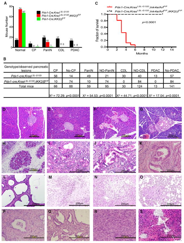
(A) Numbers of mutant mice that developed PDAC, cystic ductal lesions (CDL), PanIN, or chronic pancreatitis (CP), or remained healthy, in cohorts of Pdx1-Cre;KrasLSL-G12D, Pdx1-Cre;KrasLSL-G12D;IKK2/βF/F, and Pdx1-Cre;IKK2/βF/F mice.
(B) Chi-square analysis of the association between Pdx1-Cre;KrasLSL-G12D and Pdx1-Cre;KrasLSL-G12D;IKK2/βF/F, and the observed phenotypes in (A). Note: CP was found in PanIN, CDL, and PDAC; PanIN was coexisted with CDL and PDAC; and CDL was observed in PDAC.
(C) Kaplan-Meier PDAC-free survival curve for Pdx1-Cre;KrasLSL-G12D;Ink4a/ArfF/F (n = 16) and Pdx1-Cre;KrasLSL-G12D;IKK2/βF/F;Ink4a/ArfF/F mice (n = 15). According to the approved animal protocol, mice that presented in a moribund state were killed for autopsy.
(D–S) Representative pancreatic histologic views. (D) Normal pancreas from a wild-type mouse. (E) Histologic appearance of normal pancreas from a Pdx1-Cre;IKK2/βF/F mouse. (F) Histologic appearance of normal pancreas from a Pdx1-Cre;KrasLSL-G12D;IKK2/βF/F mouse. (G) A rare PanIN-1 from Pdx1-Cre;KrasLSL-G12D;IKK2/βF/F mouse. (H–O) Pdx1-Cre;KrasLSL-G12D mice. (H) Chronic pancreatitis (I) PanIN-1. (J) PanIN-2. (K) PanIN-3. (L) Cystic ductal lesion. (M) PDAC. (N) PDAC liver metastasis. (O) PDAC lung metastasis. (P-R) Pdx1-Cre;KrasLSL-G12D;Ink4a/ArfF/F mice. (P) PanIN. (Q) Cystic ductal lesion. (R) PDAC. (S) Histologic appearance of normal pancreas from a Pdx1-Cre;KrasLSL-G12D;Ink4a/ArfF/F;IKK2/βF/F mouse. See also Figure S1.