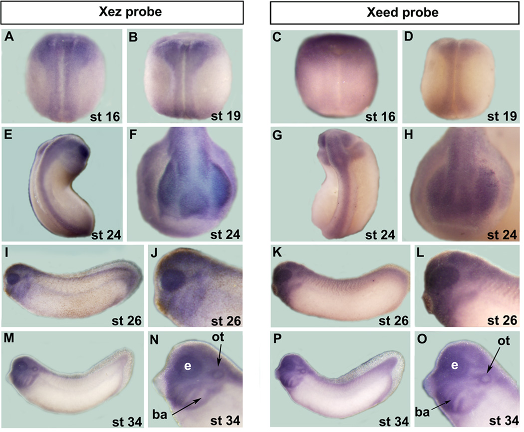
Fig. 4.
Comparison of Xez and Xeed spatial expression during frog development as assessed by whole mount in situ hybridization. A B, C, D, E and G are dorsal views. F and H are anterior views. I, K, M and P are lateral views. J, L, N and O are magnifications of the head region in I, K, M and P, respectively. ot, otic vesicle; ba, branchial arches; e, eye.

