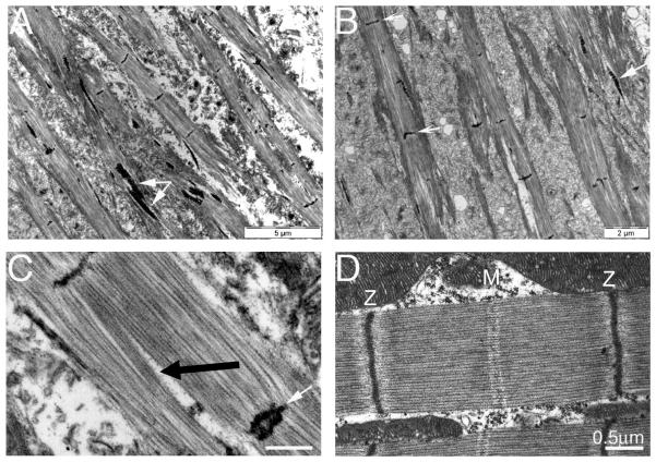
Figure 2.
Electron micrographs of flight muscle sarcomeres from Act88F-G15R homozygous mutant (A-C), and wild type flies (D). In panel A and B aberrant myofibrils are seen; there is a lack of regular structure; Z-discs never extend across the myofibril width and large arrays of ‘zebra-bodies’ comprising stacks of short sections of Z-disc-like material, spacing by about 200nm, are seen mostly within the muscles. In this and other mutants they are also found separated from the myofibrillar materials. Panel C highlights other aspects of these mutant sarcomeres including longitudinal gaps (black arrow) and dark staining material which appears to be split or duplicated Z-discs (white arrow). Panel D, wild sarcomere shows the very regular structure and much reduced I-band width, typical of this muscle type. EMs courtesy of Vikash Kumar.

