Fig. 4.

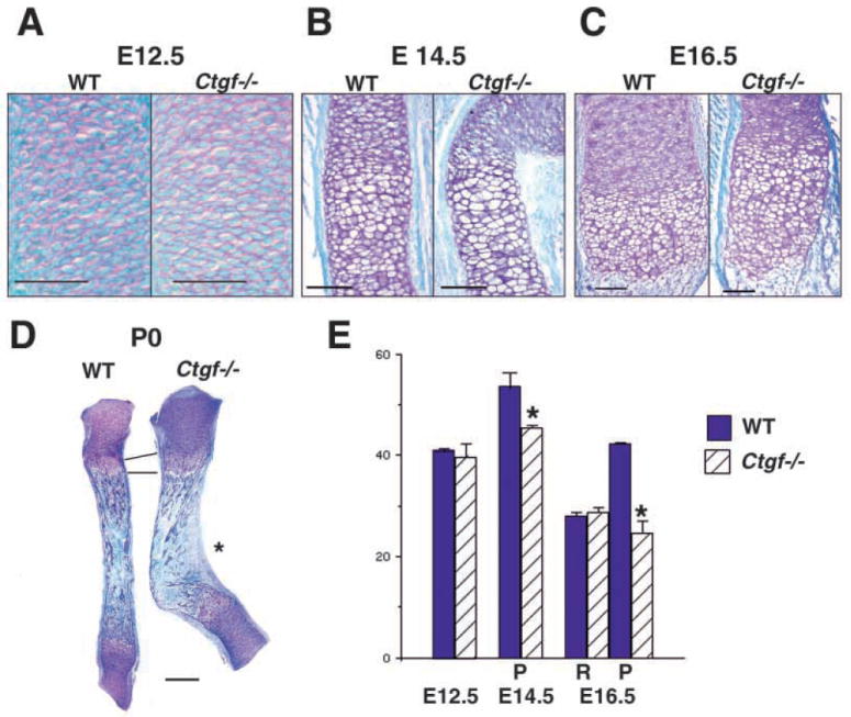
Histological and proliferative defects in Ctgf mutant cartilage. (A) Sections through wild-type and Ctgf mutant humeri at E12.5, showing no apparent differences in size or morphology. Scale bar: 50 μm. (B) Sections through wild-type and Ctgf mutant E14.5 radii at the metaphysis. Hypertrophic cells are present in wild-type and mutant littermates. Scale bar: 100 μm. (C) Sections through growth plates of E16.5 wild-type and Ctgf mutant radii demonstrate that the growth plate is expanded in mutants. The junction between the zones occupied by prehypertrophic and hypertrophic chondrocytes is disorganized in mutants. Scale bar: 100 μm. (D) Radii from newborn wild-type and Ctgf mutant littermates, demonstrating persistence of the enlarged hypertrophic zone. The epiphyses appear normal in mutants. The concave surface of the kink in mutants is a site of membrane bone formation (asterisk). Scale bar: 300 μm. (E) Reduced rates of chondrocyte cell proliferation in Ctgf mutants. Quantification of PCNA labeling is shown in the graph, with values expressed as % labeled nuclei. Cells in five adjacent sections, each spanning 40 μm, were scored by an observer blinded to the genotype. Statistical significance was assessed by Student’s t-test. *P<0.01; P, proliferative zone; R, resting zone.
