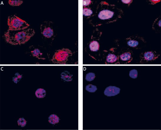
Figure 2.
Binding of anti-Dsg-3 autoantibodies to HaCaT analysed by confocal microscopy. Cells grown directly on tissue culture chamber slides were incubated with anti-Dsg-3 autoantibodies isolated from PV patients in the active stage (A) or in remission (B) used at 1 mg/ml. Control cells were incubated with IgGs obtained from healthy relatives (C) of control donors (D) also used at 1 mg/ml. Then they were rinsed with PBS, fixed with 3% paraformaldehyde for 20 min on ice, blocked with 10% normal goat serum and after blocking with BSA stained with goat antihuman IgG conjugated with TRITC. Cell nuclei were visualized using Hoechst 33342

