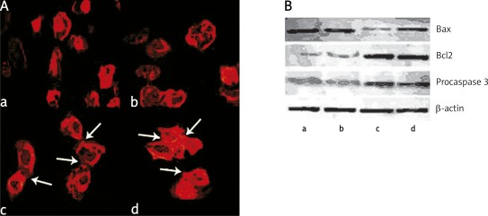
Figure 3.
Reorganization of cytoskeleton induced by anti-Dsg-3 autoantibodies. HaCaT cells were incubated for 24 h with anti Dsg-3 autoantibodies isolated from patients in active (panel A, picture a) or remission stage (panel A, picture b) used at the concentration of 1 mg/ml. Control cells were incubated with IgG of healthy relatives (picture c) or control donors (picture d) used at the same concentration. Then, HaCaT cells were rinsed with cold PBS, dipped in −20°C methanol for 20 min and stained with Texas Red-X phalloidin to visualize actin filaments. Panel B shows Western blot analysis of Bax, Bcl proteins and procaspase 3 levels in HaCaT cells incubated in the presence of anti Dsg-3 autoantibodies isolated from PV patients at the active stage (lane a), patients in remission (lane b), healthy relatives (lane c) and healthy donors (lane d). β-Actin was used as a control for SDS/PAGE loading

