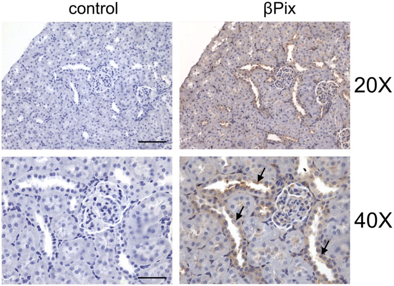
Figure 6.
βPix expression in rat kidney tissue. Representative immunohistochemical staining for βPix detection (right) in kidney cortical sections of Sprague-Dawley rat. Original magnifications, ×20 (upper; scale bar: 100 μm) and ×40 (lower; scale bar: 50 μm; cortical collecting ducts (CCD) are shown by arrows. Reproduced from Pavlov et al., 2010 with permission).

